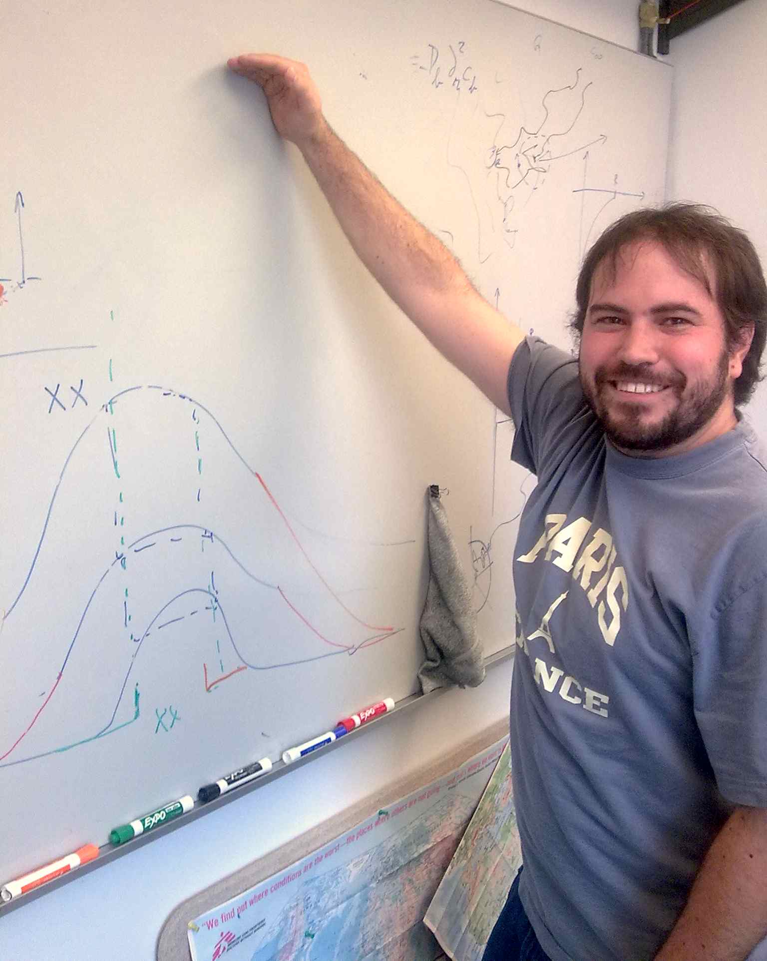
Ignacio Calderon
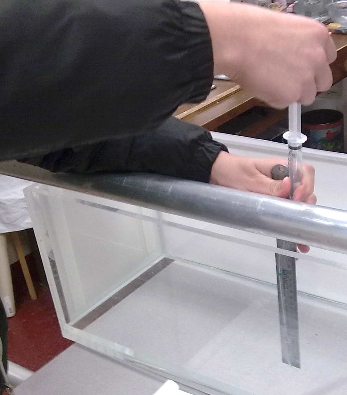
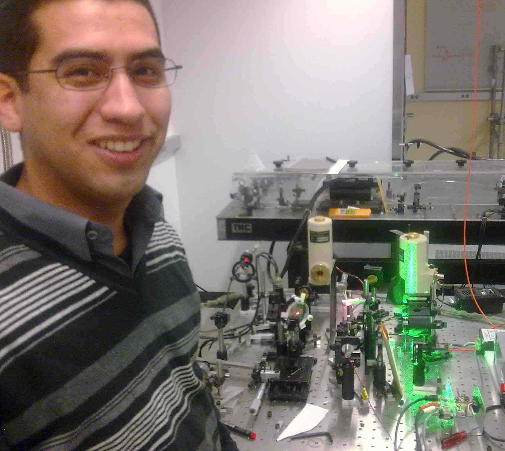
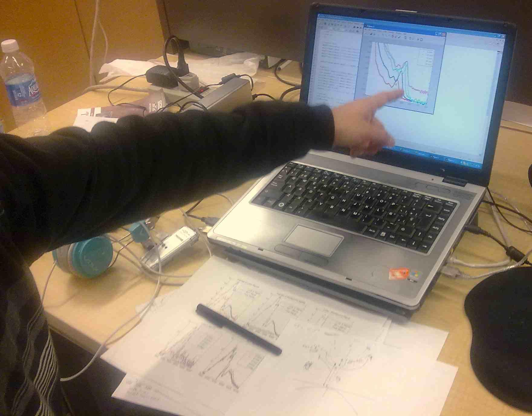
Ignacio is studying surface layers made with nanoparticles 15 nanometers across in the research group of Dr. Binhua Lin. When placed on the surface of water, these nanoparticles make an elastic skin. Ignacio measures the stresses in this skin using the Langmuir trough at top. Two strips of paper are hanging into the trough. One can see the deformed meniscus from the strip at the left. By measuring the force on the two hanging wires, one can determine the stresses in the two diretions. At right Ignacio shows his stress data: stress (vertical axis) grows as the surface area (horizontal) decreases. The measurement protocols Ignacio is developing will help future studies of nanolayer elasticity. --15Feb2011
Sergio Correa
|
 |
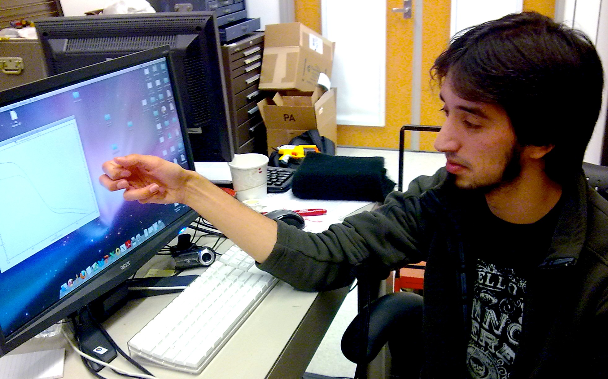
Cristian Fernandez
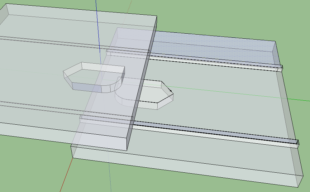
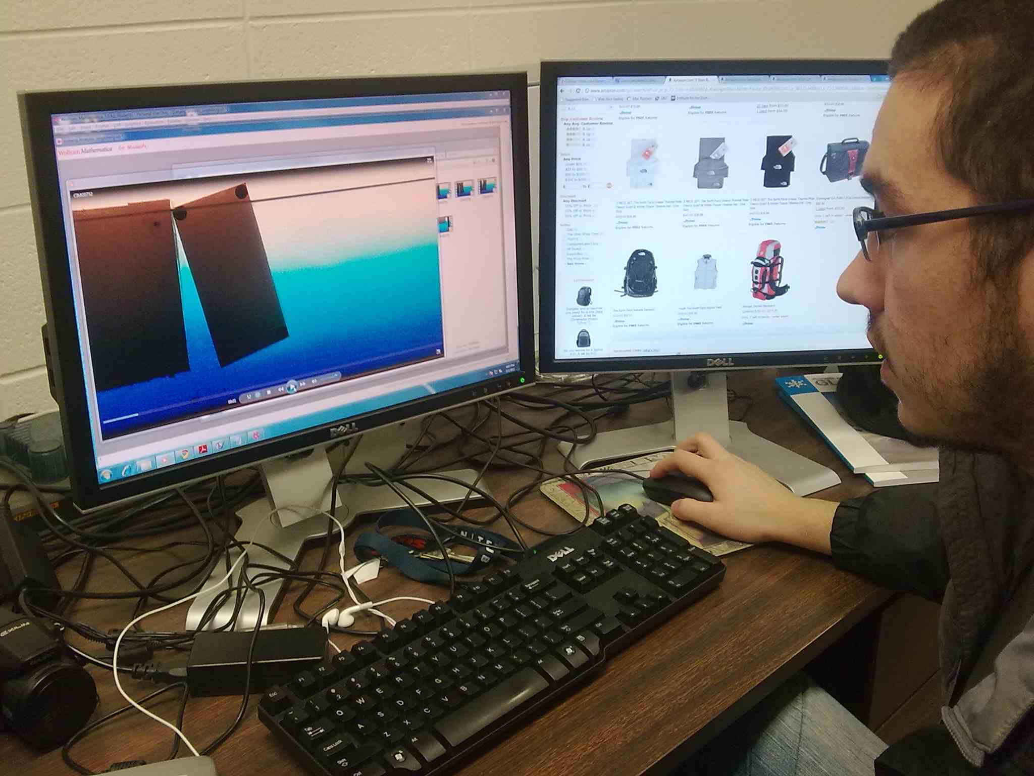
Cristian is working with Prof. Witten to understand a fluid breakup phenomenon unearthed in Prof. Ismagilov's lab. The sketch at the left shows two microscope slides with a tiny well etched into each one. Water (not shown) is held in the wells by capillary action. When the the wells slide apart, the water makes a transition from one drop to two. Cristian's numerical calculations show the shape of the water near the moment of transition where the water divides. He studies the case of two right-angle corners sliding apart, like the rear edge of the wells in the sketch. Here it appears that the transition is continuous, contrary to expectations. Cristian's sketch at right attempts to account for this continuous shape change.
Andres Franco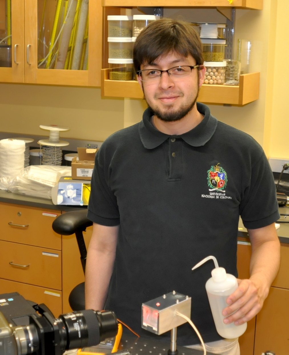
Andres is working with postdoc Justin Burton in Prof. Nagel's lab on the motion of a floating Leidenfrost drop. The dancing droplet of water flicked onto a hot skillet is a Leidenfrost drop. Andres's setup is shown in the center picture. The drop is just visible at the top; a magnified image of it appears in the glass prism below. It is not flat on the bottom; the water evaporating from the bottom surface pushes it upward to form a convex bulge. The drop comes closest to the heating surface in a thin, ringlike rim about 100 microns deep.
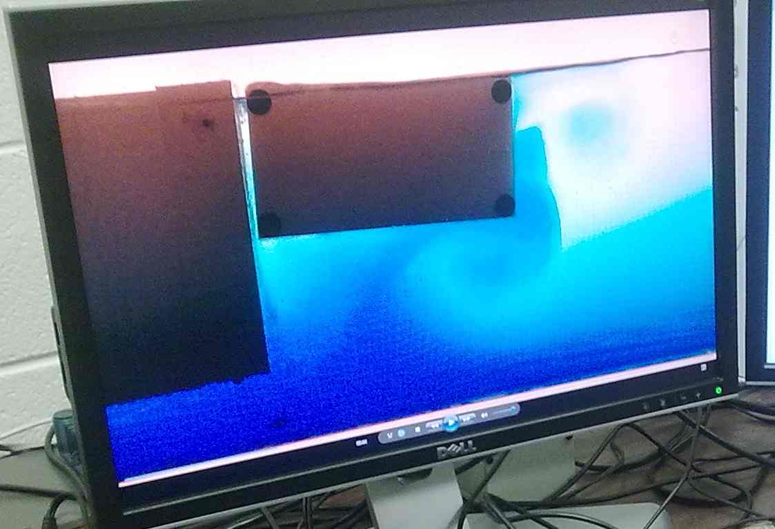
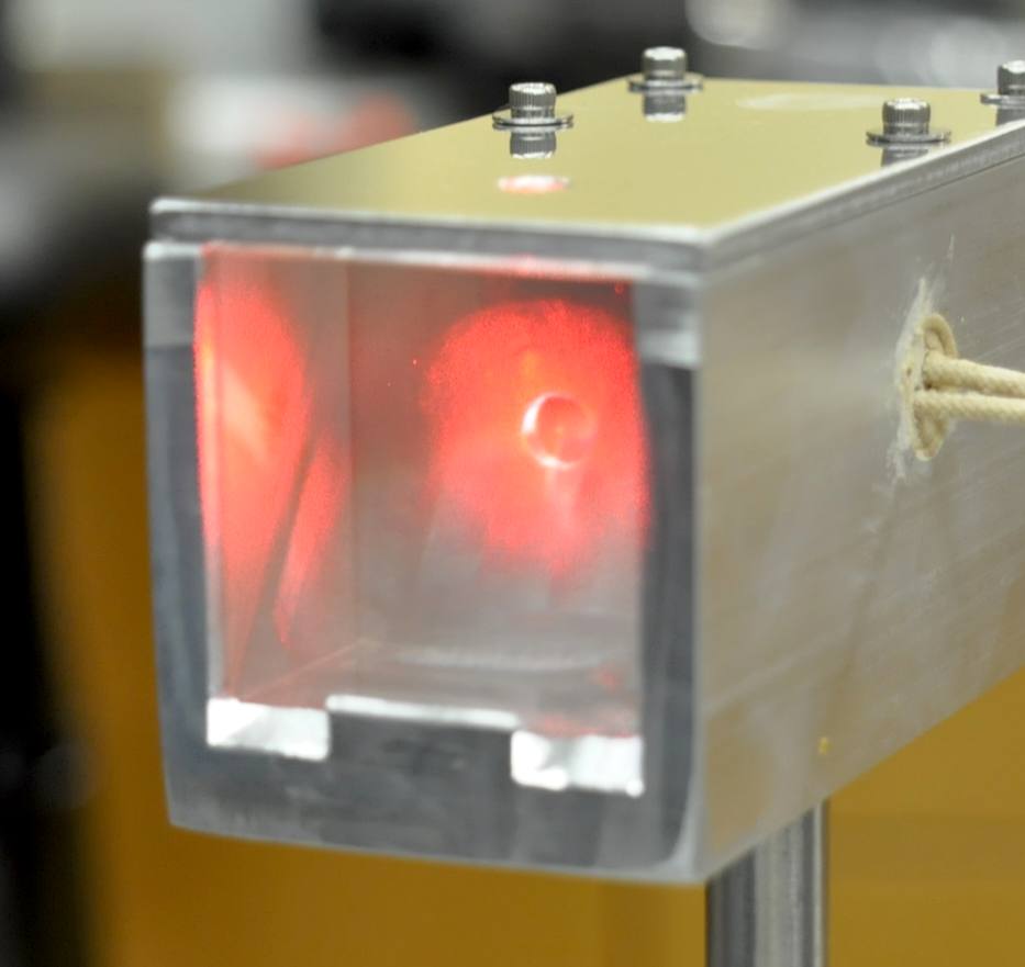
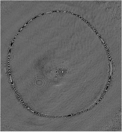
The question of interest is how the liquid is moving in this thin ring with gas rushing outward across it. To find out, Andres and Justin shine a laser beam on the drop from beneath and observe the interference fringes from light reflecting from the liquid. The interference pattern is shown at right.
The fringes move rapidly. to capture the motion they use a fast camera that captures a picture every 30 microseconds. The gas passing under the ring excites capillary waves. Now they want to determine the thickness profile from the moving fringe patterns. They want to see the amplitude of the capillary waves, and which direction they move as a function of the strength of heating..
Roberto Gomez
Carolina Perez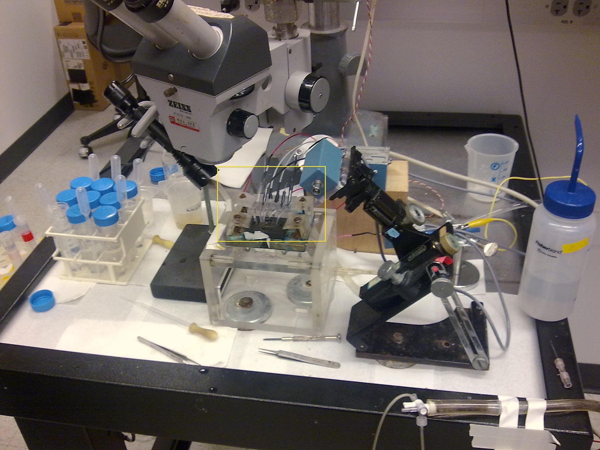
Carolina's subject is ion channels, the molecular valves that control the activation of muscles, nerves and other cells in the body. Working in Prof. Francisco Bezanillo's lab, Carolina is trying to slow down the controlling movement of the channel by fastening parts of the constituent proteins together. The native protein molecules are modified at two points by altered amino acids that bind the two points together. This modification is done genetically, by alternating the DNA instruction code for these proteins. The modified DNA is injected into frog oocytes (eggs). Then the normal functioning of the cell creates the modified protein instead of the native protein. The oocyte membrane is coated with the modified ion channels.
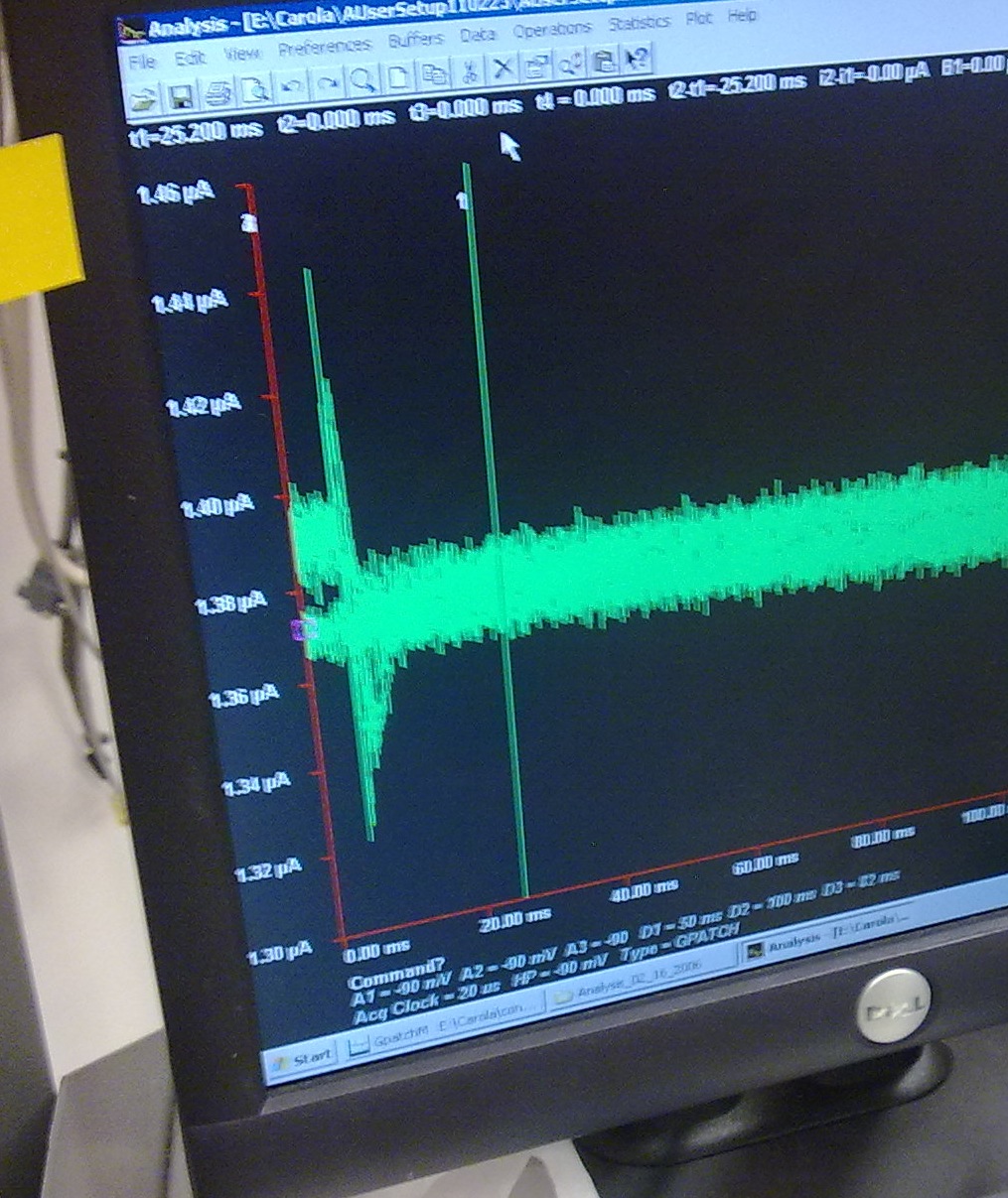
The channels are then tested by measuring their electrical response. A section of the oocyte membrane is placed across a hole in a thin sheet of glass or plastic. Electrodes are then placed on either side of the channels. By controlling the voltage across the membrane and the current through it, one can infer how long the channels stay open after a stimulus opens them. The membrane and electrodes are packaged into the device at right marked by the yellow box. A typical time trace of the current is shown at left. One sees a downward spike at left that returns to the baseline as one moves to the right.
Estefania Vidal
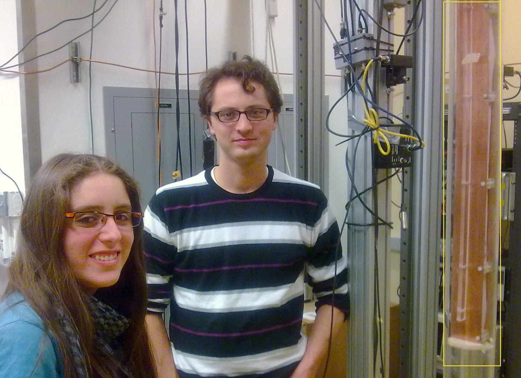
 Estefania and her
grad-student mentor Scott Waitukaitis (a former
intern in the program) are next to Scott's experiment to measure grains
falling an electric field in the lab of Prof. Heinrich
Jaeger One copper sheet electrode of
the
falling chamber is indicated by the yellow box. Somehow the
collisions of
the grains gives them charges, both positive and negative.
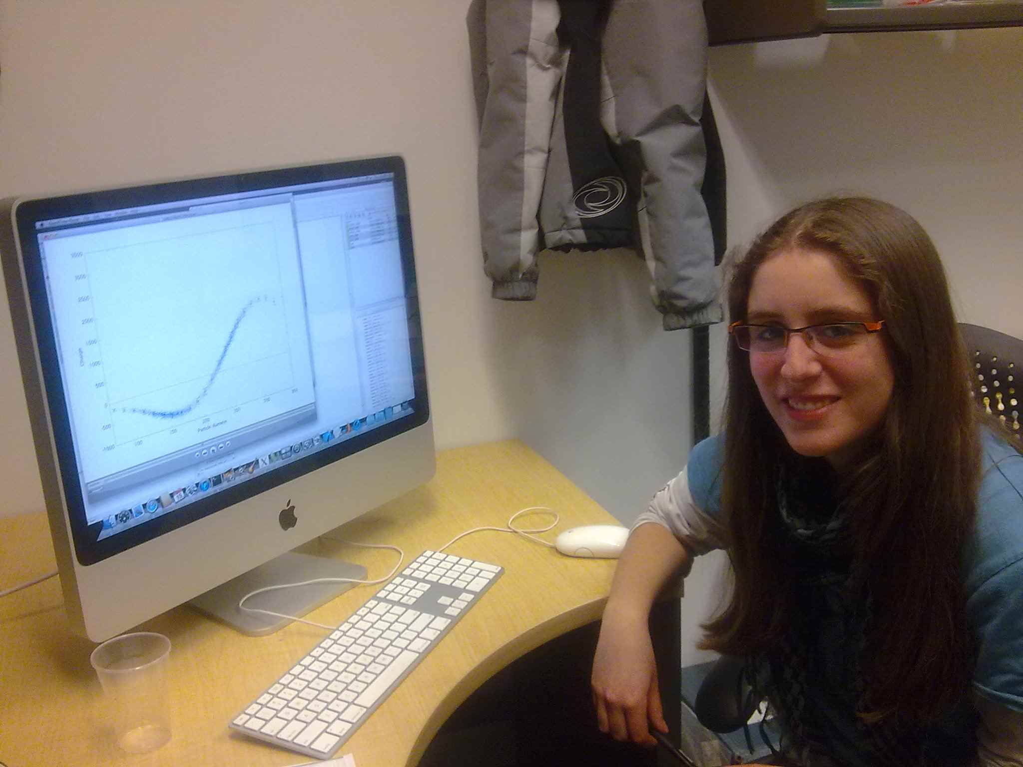
Estefania's job is find how the probability distribution of the charge
by a computer simulation that follows each grain through hundreds of
collisions with other grains. Her data on the screen at right
gives a good match to Scott's experimental measurements.
--- 16Feb2011
Estefania and her
grad-student mentor Scott Waitukaitis (a former
intern in the program) are next to Scott's experiment to measure grains
falling an electric field in the lab of Prof. Heinrich
Jaeger One copper sheet electrode of
the
falling chamber is indicated by the yellow box. Somehow the
collisions of
the grains gives them charges, both positive and negative.
Estefania's job is find how the probability distribution of the charge
by a computer simulation that follows each grain through hundreds of
collisions with other grains. Her data on the screen at right
gives a good match to Scott's experimental measurements.
--- 16Feb2011