Research
Force Transmission at Cell Adhesions:
Forces generated within the actin cytoskeleton are transmitted to the extracellular matrix at integrin-mediated focal adhesions and neighboring cells at cadherin-mediated cell-cell contacts. Our goal is to delineate the mechanical feedback between actomyosin forces, cell adhesion and extracellular mechanical cues by using a combination of traction force microscopy and live cell imaging approaches. Recently, we have shown that force transmission during myosin-II mediated focal adhesion maturation is modulated through tension-dependent “clutch” between transmembrane integrins and the extracellular matrix (Aratyn-Schaus and Gardel, 2010). Current reviews of this area can be found in: current understanding of this field in (Aratyn-Schaus and Gardel, 2008) and (Gardel et al., 2010).

Figure (right): Forces at Cell-Cell contacts. A pair of MDCK cells expressing GFP-cadherin with traction stress vectors overlayed (red vectors). The cell pair is delineated by the green outline; a single cell delineated by the yellow. By examining the force balance across this cell pair, we can measure the forces exerted at the cell-cell contact.
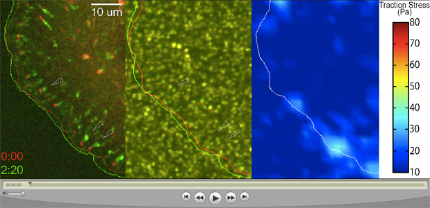
QuickTime Movie: Focal Adhesion Mobility at Low Tension (to view click on the image above). Color combined time-lapse movie of mApple-paxillin (left) and beads within the underlying substrate (middle) at successive times post washout of a myosin II inhibitor (red) and at 2:20 (green). Cell-edge boundary shown for the same times and colors. Arrows point to examples of slipping focal adhesions and corresponding regions in underlying beads. Color-coded traction stress map and scale (right) with boundary of cell-edge in white over same times post blebbistatin washout. Time is indicated in min:sec and 0:00 indicates first image post blebbistatin washout.
See also:
Self-Assembly and Biophysical Properties of Actin Networks and Bundles
| Margaret Gardel's Office
University of Chicago |
Laboratory
University of Chicago |