Research
Cellular Force Transmission:
In cells, the F-actin cytoskeleton is responsible for much of the forces generated and transmitted across individual cells; the regulation of force transmission guides cell shape change, adhesion and the degree of tension sustain on the extracellular environment. These biophysical behaviors play an important role in cell adhesion, migration and division. We seek to understand the regulation of self-assembly and force transmission through the F-actin cytoskeleton and examine the role of motor proteins and cross-linking proteins in regulating this force-dependent architecture.
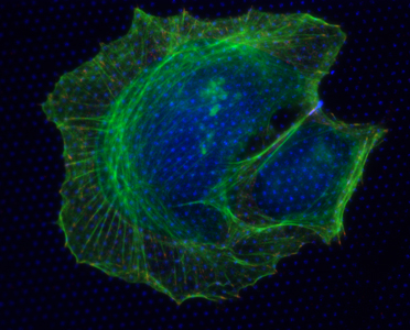
Figure (right): U2OS osteosarcoma cell plated on a substrate with micropatterned fibronectin (blue dots) to constrain the location and size of focal adhesions, visualized by paxillin (red), and the organization of F-actin (green).

QuickTime Movie (above). To view click on the image. Reorganization of actomyosin cytoskeleton and traction stresses after removal of the myosin II ATPase inhibitor, blebbistatin. Spinning Disk confocal time-lapse movie of GFP-actin (left) and mApple-paxillin (middle) prior to and after blebbistatin treatment and removal. Color-coded traction stress map with cell-edge outline in white (right). This cell was plated on a 2.8kPa PAA gel. Time is indicated in min:sec and 0:00 indicates first image post blebbistatin washout.
See also:
Force Transmission at Cell Adhesions
Self-Assembly and Biophysical Properties of Actin Networks and Bundles
| Margaret Gardel's Office
University of Chicago |
Laboratory
University of Chicago |